
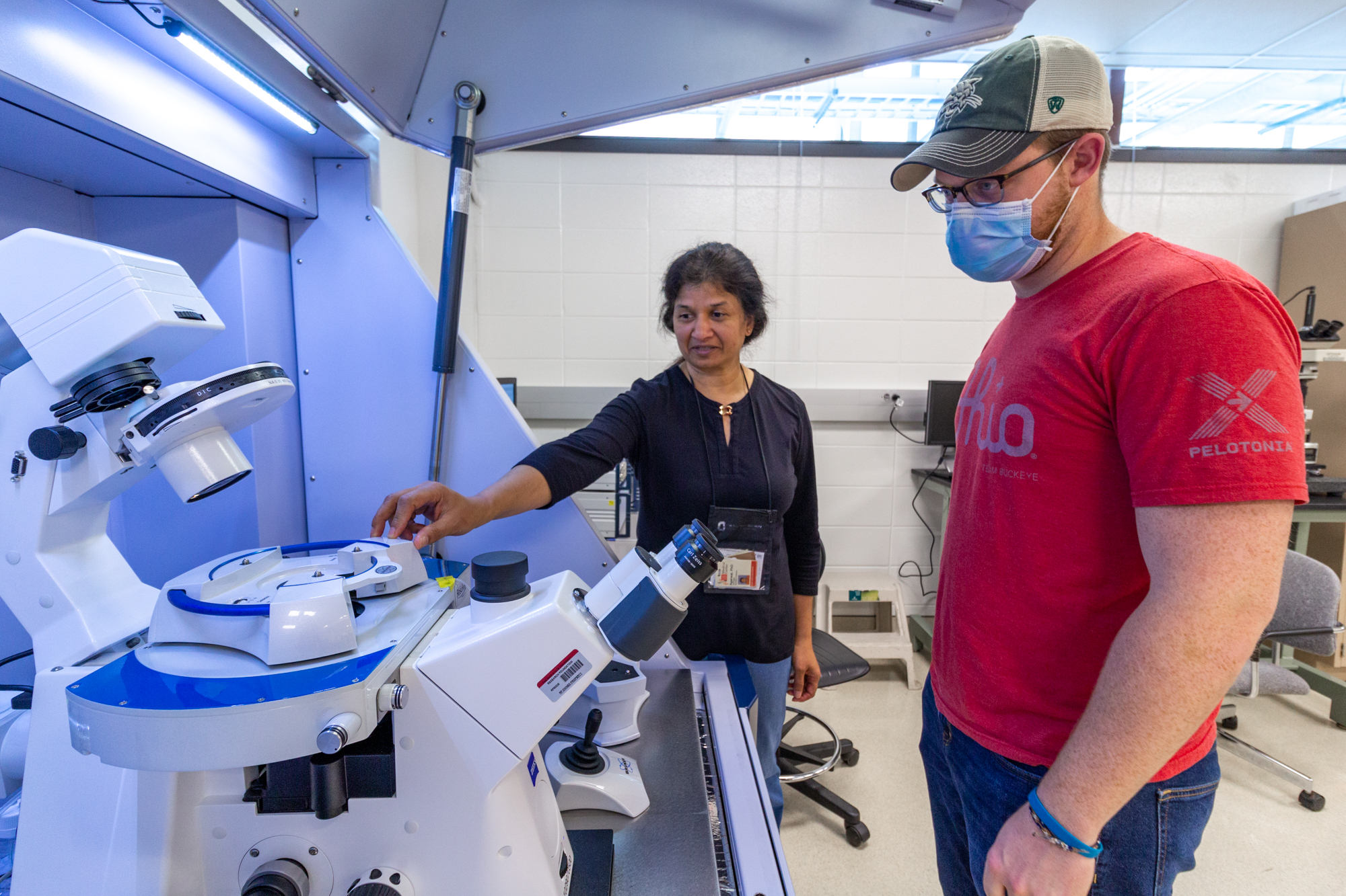
Gunjan Agarwal, left, and a student with the AFM in Scott Lab.
A newly installed atomic force microscope (AFM) coupled to a fluorescence light microscope is open for shared use in Scott Laboratory W374 (A/B).
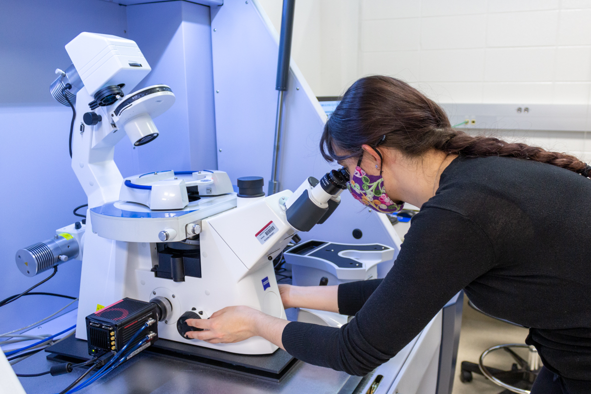
This multi-user facility is home to a state-of-the-art Bruker Resolve AFM, enabling novel applications in imaging, analyzing and manipulating matter at the nanoscale. Atomic force microscopy enables the imaging and nano-mechanical analysis of most types of surfaces, including polymers, ceramics, and biological samples.
“The nanoscale and molecular-level insights that can be provided by AFM are expected to generate new research directions for basic and translational biomedical research as well as in nanotechnology and polymer science,” said Gunjan Agarwal, professor in Mechanical and Aerospace Engineering (MAE) who leads the lab. “This AFM can analyze samples in a liquid environment at the nanoscale level, which cannot be achieved by other existing AFMs on campus or by electron microscopy.”
 The microscope was acquired via a National Institutes of Health (NIH) S10 award, led by Agarwal. This is the second S10 award led by Agarwal, who had previously established and directed another Bio-AFM facility in the Davis Heart and Lung Institute, in 2006.
The AFM laboratory as a university-wide shared resource will enable collaborations across various departments and colleges, as can already be witnessed in some of the user projects.
The Institute for Materials Research (IMR) is set to provide the lab further support through its first five years of operation. Along with IMR, the Office of Research, departments of MAE and Biomedical Engineering, and the colleges of Engineering, Arts and Sciences, and Pharmacy have committed funds to support this facility.
The new AFM system offers unique capabilities to users at Ohio State.
First, it is the only shared AFM on campus capable of imaging in fluid, making it an invaluable resource for biomedical research.
“In biological samples, it is really important that we can keep the samples hydrated (e.g. live or fixed cells, tissues and even biomolecules) and in their desired conditions,” Agarwal said. “The instrument is very bio-friendly.”
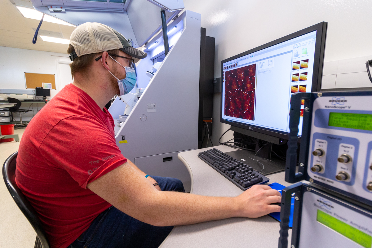 Additionally, the instrument’s AFM module sits above a light microscope, allowing the coupling of fluorescence and AFM images from the same region. In the examination of bio samples, fluorescent signals will allow users to perform AFM analysis of specific regions of interest identified in fluorescence images.
Finally, the new AFM is equipped with state-of-the art features like the PeakForce Tapping mode, Quantitative Nano mechanics, and even Kelvin Probe Microscopy.
Agarwal has also developed and offers a new course MECHENG 6194 (ME 6711) “Microscopy in Biomechanics” that discusses the fundamental principles of light microscopy and AFM in imaging and nanoscale mechanical analysis.
Story by Mike Huson, IMR Communications Coordinator
Contact: huson.4@osu.edu
Follow: @OhioStateIMR